SP 5
Image based modelling (DHZB/Charite)
Image based modelling offers the unique opportunity to assess and simulate (predict) the individual patient’s response to treatment and to link clinical information of function, tissue composition and anatomy with modelling at various scales.
Objectives
• The goal of this WP is to provide detailed simulation models of patient’s hemodynamics and cardiac biomechanics in response to treatment and to combine this information in an iterative fashion with the other modelling modalities of SMART.
• Imaging will be based on MR imaging and derived data will be used as model inputs. This includes: global pump function (chamber size, ejection fraction, myocardial mass), fibrous tissue content, 4D blood flow in the ventricles and the aorta (, myocardial wall motion and deformation. The key-parameters that will be modelled for the simulation of treatment effects (outputs of modelling) are myocardial deformation, pump efficiency and kinetic energy loss of blood.
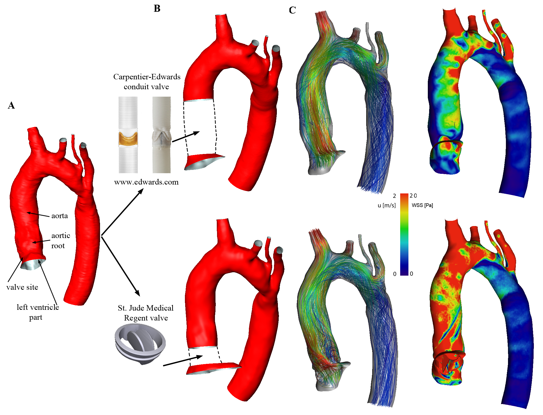
• Virtual valve replacement of mechanical and biological valves will be accomplished using Computed Aided Design software. A set of commercially available valves, that will be used at the DHZB, will be scanned optically with a resolution of 0.1 mm in a fully open position using an optical method.Than the valve geometry will be incorporated into the anatomical models of the individual patients.
Tasks
• Image acquisition methods include (a) cine MRI (ventricles and aorta), (b) 4D blood flow (ventricles and aorta), ( (c) T1-maps of the ventricles.
• Bioimaging data extraction and postprocessing will be done by an extended CAIPI framework (www.mevis.fraunhofer.de/loesungen/cardiac-mri-inspection-of-the-heart-muscle.html) that allows for advanced integration and analysis of CV imaging data form research and clinics. The current framework already allows the integrated 4D analysis of heart motion, myocardial tissue characterization and blood flow. As an efficient, highly parallelizable flow solvers the Lattice-Boltzmann Method is used.
• Patient-individual anatomical and functional models are created, which can simulate local changes in myocardial motion and hemodynamics. The work includes motion analysis with phase-based registration, segmentation of heart chambers and myocard.